Phone:(207) 481-8232
Charlie’s Journey With an Oral Plasmacytoma
A fast-growing oral mass led Charlie to our dental team, where advanced imaging, biopsy, and jaw surgery helped give her a positive outcome.
A fast-growing oral mass led Charlie to our dental team, where advanced imaging, biopsy, and jaw surgery helped give her a positive outcome.
Case of the Month: Meet Charlie!
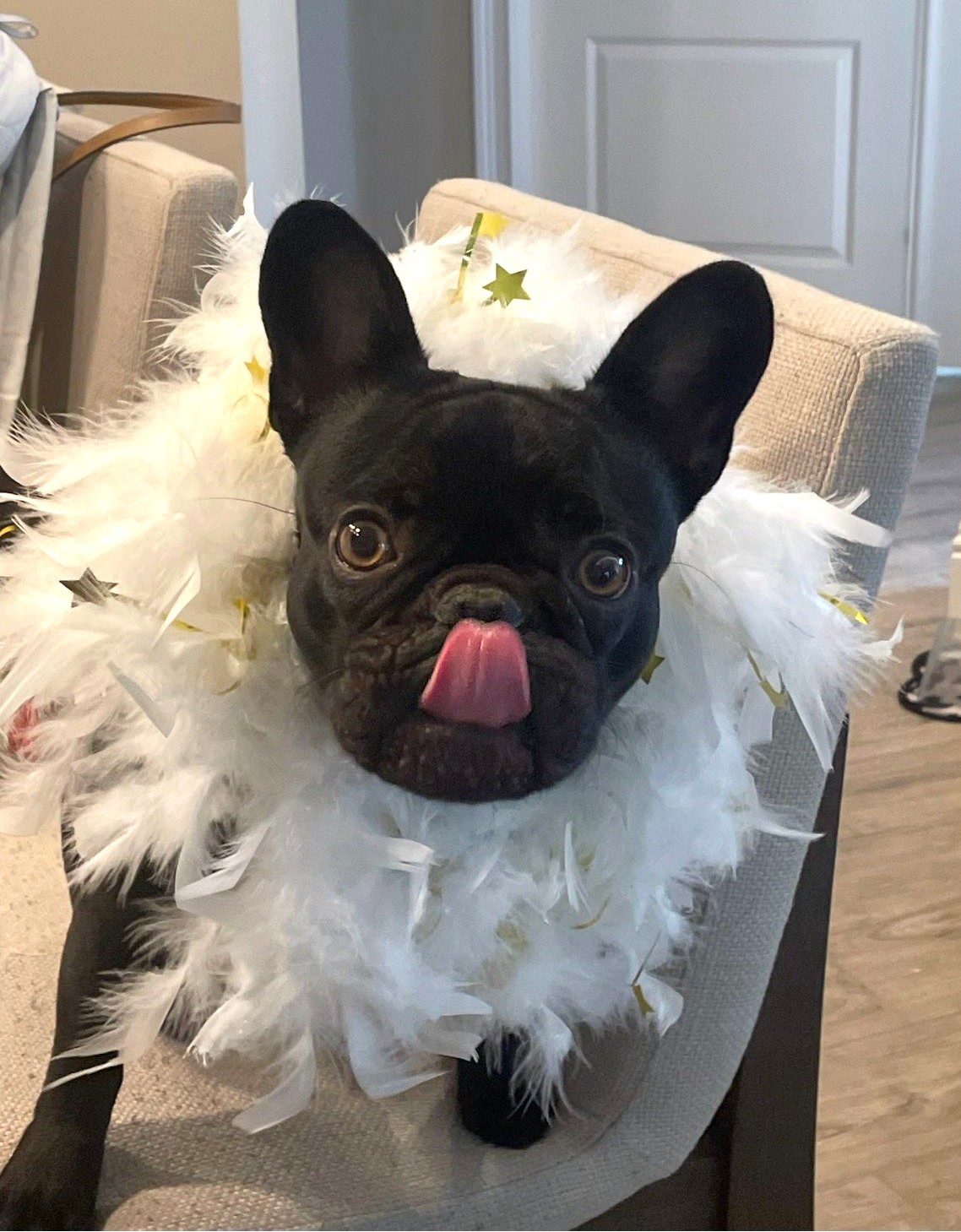
Charlie is a 2-year-old French Bulldog who came to Mainely Veterinary Dentistry & Urgent Care for a large, fast-growing mass on the lower left side of her mouth.
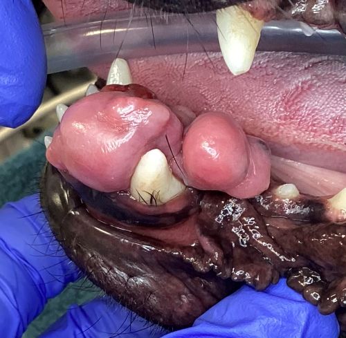
Her family had noticed tissue growing around several of her teeth and pushing her lower front teeth toward the right. The area would also bleed, and Charlie had developed bad breath that temporarily improved with antibiotics before returning.
After Charlie’s primary veterinarian became concerned that the growth might be more than simple gum overgrowth, her family was referred to our veterinary dental team for further evaluation.
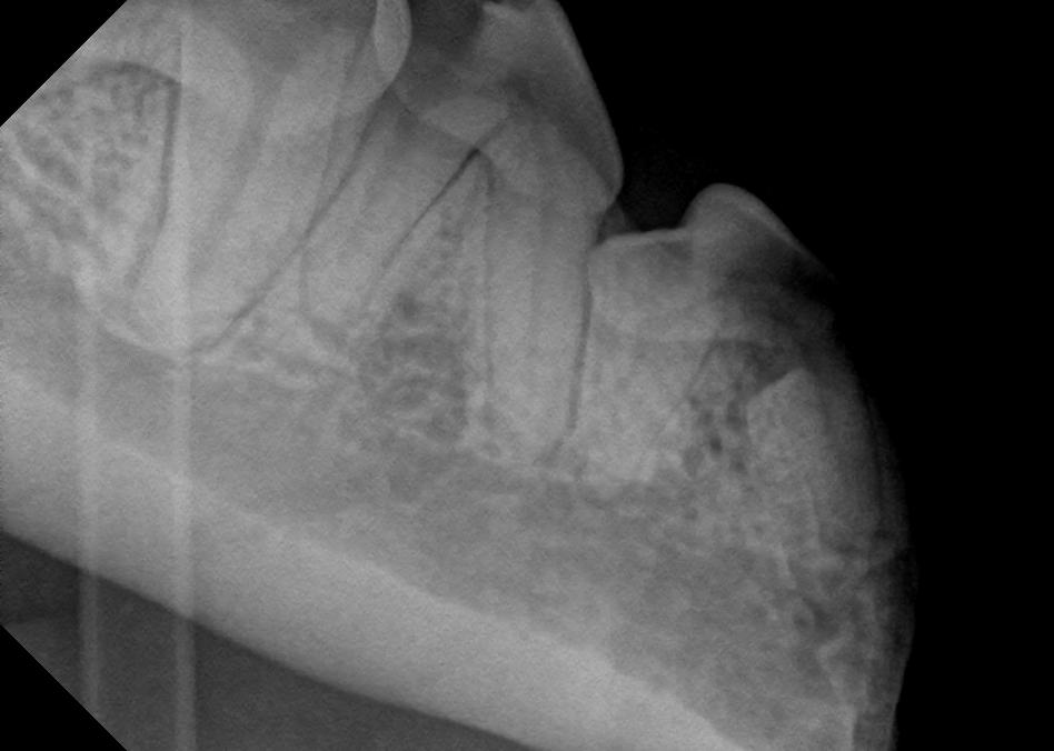
During Charlie’s oral examination, Dr. Jennifer Keaten found a large mass surrounding her lower left canine and incisor teeth.
Before deciding how to treat it, we needed to know exactly what type of mass we were dealing with and how extensively it involved the surrounding structures.
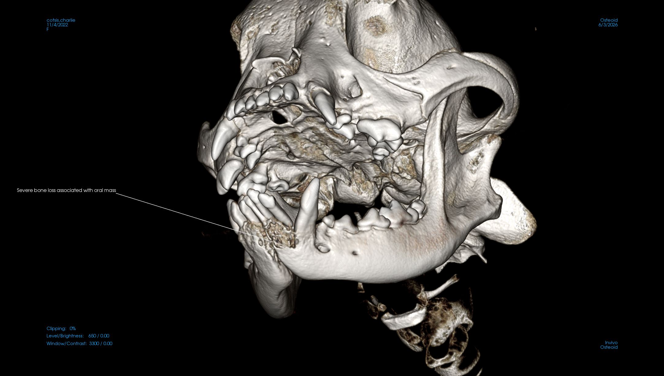
Charlie returned just a few days later for a Cone Beam CT scan and biopsy. The 3D imaging allowed our team to evaluate the mass and underlying jaw in much greater detail, while the biopsy provided tissue that could be examined by a veterinary pathologist.
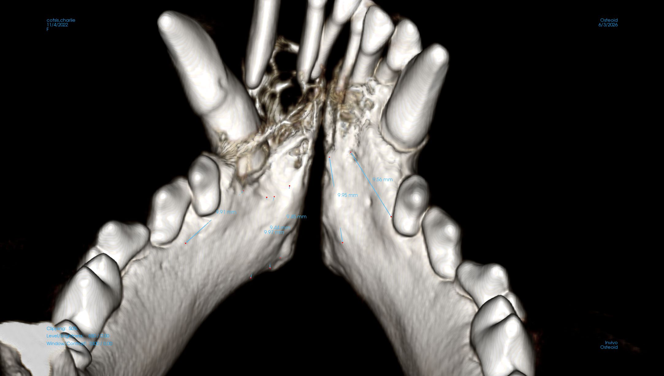
Charlie’s biopsy was submitted to Specialty Oral Pathology for Animals (SOPA), where it was evaluated by Dr. Cindy Bell, who specializes in oral pathology.
The diagnosis was an oral plasmacytoma.
A plasmacytoma is a tumor made up of plasma cells, a type of immune cell involved in producing antibodies.
Oral plasmacytomas can grow rapidly and may invade nearby tissues, including the underlying bone. Fortunately, spread to distant parts of the body is uncommon, and surgical removal can provide a very good outcome for many dogs.
These tumors are generally seen in older dogs, making Charlie’s diagnosis at only two years old particularly unusual.
Her case is an important reminder that age alone cannot rule out an oral tumor. A new or changing growth in a pet’s mouth should always be evaluated.
Once we knew the diagnosis, we could develop a surgical plan designed to remove the entire mass with an appropriate margin of surrounding tissue.
For Charlie, that required a rostral mandibulectomy, a procedure that removes a portion of the front of the lower jaw.
We also recommended chest X-rays and an abdominal ultrasound to look for any evidence that the tumor had spread and to identify other health concerns that could affect anesthesia or treatment.
Thankfully, there was no evidence of metastasis.
The chest X-rays did reveal a likely sliding hiatal hernia, a condition that can occur in French Bulldogs and may contribute to signs such as regurgitation or nausea. Knowing this ahead of time allowed our team to plan appropriately for Charlie’s anesthesia and recovery.
Four weeks after her initial examination, Charlie returned for surgery.
Dr. Keaten performed the rostral mandibulectomy, removing the affected portion of Charlie’s lower jaw along with the oral mass. The tissue was then submitted for histopathology so the surgical margins could be evaluated for remaining tumor cells.
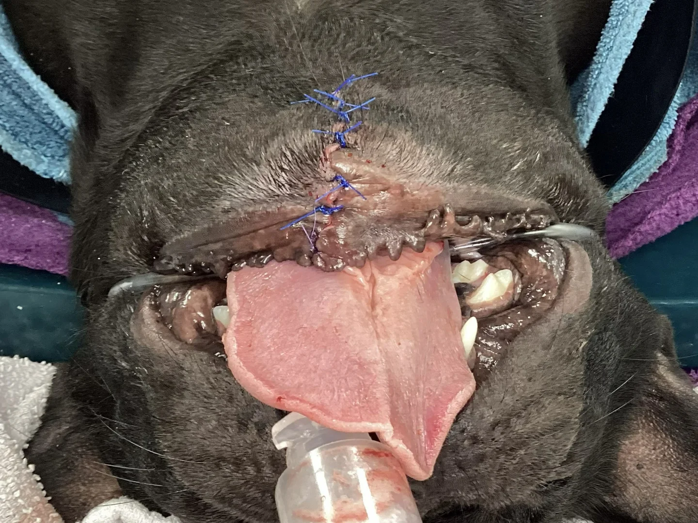
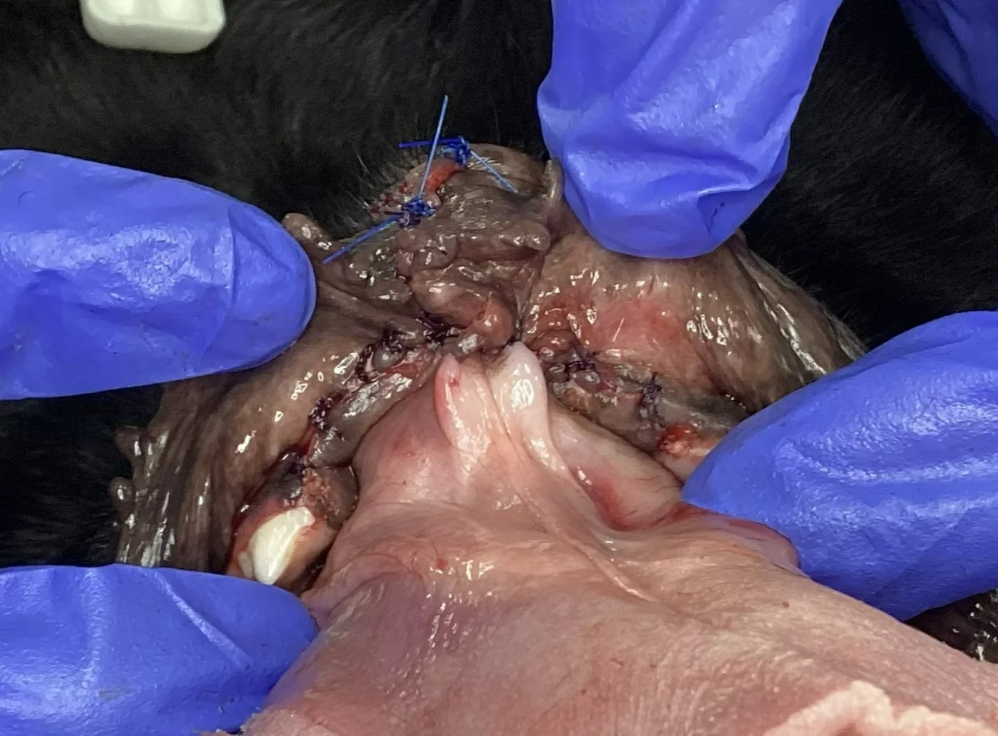
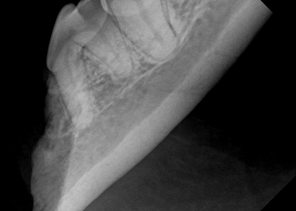
Despite how significant a jaw surgery may sound, dogs can adapt remarkably well after a partial mandibulectomy. Charlie recovered quickly, and it was difficult to tell just by looking at her that a portion of her lower jaw had been removed.
Her final pathology confirmed the plasmacytoma diagnosis. One very small area of the surgical margin may have contained a few remaining plasmacytoma cells, although additional bone had been smoothed away before the surgical site was closed.
Charlie’s family is considering whether additional radiation therapy is appropriate. For now, the plan is to monitor the area closely for any sign of the tumor returning and reconsider radiation if recurrence is detected.
Oral tumors do not always cause obvious symptoms when they first develop. Changes worth having evaluated include:
These signs do not necessarily mean that your pet has cancer. Infections, periodontal disease, benign growths, and other conditions can cause similar changes. An examination and, when appropriate, imaging and biopsy are needed to determine the cause.
Charlie’s case is also a reminder that veterinary dentistry is about much more than cleaning teeth.
A thorough oral examination provides an opportunity to identify abnormal tissue, damaged teeth, periodontal disease, and other changes that pet owners may not be able to see at home.
Our team recommends beginning routine professional dental care early in a pet’s life and continuing regular oral evaluations over time. If you notice a lump, bleeding, unusual odor, loose teeth, or another change in your dog or cat’s mouth between visits, it is best to have it evaluated rather than waiting for the next routine cleaning.
Today, Charlie is back to enjoying life with her family and has recovered beautifully from surgery. We will continue monitoring her closely, but we are optimistic about her future.
Her story shows how early evaluation, advanced imaging, biopsy, and specialized oral surgery can work together when an unexpected mass is discovered.
If your pet has an oral mass or another complex dental concern, learn more about our advanced veterinary dental services or schedule an evaluation with our team.