Phone:(207) 481-8232
Detecting Oral Masses in Cats and Dogs: Why Early Diagnosis Matters
Learn more about oral tumors in pets and understand why routine dental exams are crucial for early detection and treatment.
Learn more about oral tumors in pets and understand why routine dental exams are crucial for early detection and treatment.
Oral tumors are, unfortunately, a common concern in both cats and dogs. These masses can range from benign growths to aggressive cancers — and the earlier they’re found, the better the outcome. At Mainely Veterinary Dentistry, our use of Cone Beam CT technology during routine COHATs (Comprehensive Oral Health Assessment and Treatments) has significantly improved our ability to treat and diagnose and sometimes detect oral and other head masses in their earliest stages, when treatment is more effective and less invasive.
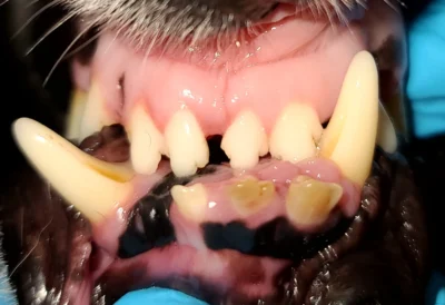
Routine dental exams are about more than just preventing tartar and periodontal disease — they’re often the only way to discover hidden masses in the mouth. Since pets don’t typically allow a full oral exam while awake, many tumors go unnoticed until they grow large enough to cause discomfort or visible symptoms.
That’s where routine annual or biannual COHATS and Cone Beam CT come in. This advanced imaging tool allows us to create detailed 3D scans of your pet’s skull, uncovering tumors or abnormalities that may not show up on traditional x-rays. When paired with a thorough oral exam under anesthesia during a COHAT, Cone Beam CT helps us identify small masses or other problems early, sometimes before they become visible to the naked eye.


Both cats and dogs can develop oral masses — some benign, others malignant, and some non-cancerous altogether (such as inflamed tissue). Unfortunately, there’s no way to determine the nature of a mass just by looking at it. That’s why we recommend surgical sampling and histopathology for a definitive diagnosis.
One of the most common benign tumors in dogs, this mass typically forms near the periodontal ligament. Some are small and easily removed, while others may require removal of the associated tooth to prevent recurrence.
Another benign tumor, but more aggressive. These often invade the surrounding bone, requiring 1 cm surgical margins for successful removal. Cone Beam CT helps us assess how deeply these masses invade the bone, improving surgical planning and outcomes.
The most common malignant oral tumor in dogs, melanomas can vary in appearance and behavior. They often spread to lymph nodes and require wider surgical margins (2 cm). Cone Beam CT is especially valuable here, helping detect early bone involvement or lymph node changes.


The most common oral tumor in cats, SCC is highly invasive and difficult to treat once advanced. It accounts for up to 80% of feline oral cancers. Early detection is key — but due to the small size of the feline oral cavity, it’s often too late by the time symptoms appear.
Routine regular dentistry paired with Cone Beam CT could increase the chance of detecting this cancer early, giving cats a better chance at treatment options. Once a cat is symptomatic for this severely painful disease it is often too late.
When it comes to oral masses, timing makes all the difference. That’s why we recommend annual or biannual COHATs for all cats and dogs — not only to keep teeth clean, but to identify issues like tumors, fractured teeth, and bone loss before they become severe.
At Mainely Veterinary Dentistry, we include Cone Beam CT imaging as part of our advanced diagnostic process. This gives us a clearer, more accurate view of your pet’s oral and facial structures, allowing us to detect hidden tumors, determine surgical margins, and create the most effective treatment plan possible.
If your pet has not had a COHAT recently — or if you’ve noticed anything unusual in their mouth — don’t wait. Early diagnosis leads to better outcomes and more treatment options.