Kelly’s Story: The transformation of a sweet old Sheltie
Kelly is a wonderful 10 year old Sheltie that came to see our dental service recently to assess and treat her terrible dental disease. One of Kelly’s owners had recently passed away and the remaining owner was unable to care for her.
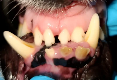
Kelly upon presentation to our dental service.
When the amazing people at the Maine Sheltie Rescue added Kelly to their rescue she was so obese that she could not support her weight on her old arthritic legs. They immediately began a diet plan and started on her journey to recovery. Anne, the wonderful founder of Maine Sheltie Rescue, also noticed there was a problem in Kelly’s mouth. Her dental disease was so significant that she had discharge coming from her nose. Anne did not waste any time and got Kelly in for a COHAT (Comprehensive Oral Health Assessment and Treatment).
It was clear from her initial oral exam that Kelly was going to need the remainder of her teeth removed due to irreversible bone loss, gum recession and inflammation. However, we did not fully appreciate the severity of her disease until we did her full oral exam and dental xrays under anesthesia and started her oral surgery. Kelly had bilateral (both sides) oral-nasal fistulas. These are holes going from the mouth into the nasal cavity. This occurs when the bone becomes infected from bacteria building up under the gumline and erodes the bone starting at the tooth root and moving eventually all the way into the nasal cavity. This most commonly occurs on the upper canine teeth as it did with our sweet girl Kelly. On the picture below to the right, the left canine tooth had fallen out on its own after many years of disease but it left the chronic hole behind into the nasal cavity. The picture on the left is before we took out the right canine tooth which revealed a much much bigger hole underneath the tooth into the nasal cavity (continue reading for a picture after extraction).
In Kelly’s case, we did not need dental xray to know that she needed the remainder of her teeth removed; however, we did need dental xray to know what the teeth looked like under the gumline to know how to extract them. Additionally, just because a tooth is missing above the gumline does not mean it is completely gone. Kelly had another very painful condition going on; she had broken one of her lower canines off under the gumline (see middle xray image below). This could have been easily missed without these important images. Finally, we also make sure to take xrays after extraction to make sure we do not leave any pieces of tooth behind.
Once we had Kelly’s upper right canine removed this is what lied beneath, a very large hole about 1 inch in diameter going straight into her nasal cavity. This was the cause for the large amount of debris coming from Kelly’s nose. Once the tooth was extracted the diseased bone was also removed and the debris was scooped and flushed from Kelly’s nose to leave a disease free and much more comfortable mouth.
After post extraction dental radiographs and preparation of the tissue for closure, the extraction site and oral-nasal fistula was closed with healing gum tissue to facilitate fast and healthy closure of the surgical areas and to prevent a chronic fistula from forming. Kelly not only received dental nerve block prior to surgery to prevent pain but also went home with pain-medication to keep her pain free during healing.
Kelly came to see us for her 2 week recheck exam and not only was her mouth feeling better and healing well but she had lost another 2.5 pounds! Kelly was much brighter and alert. She is well on her well to living out the remainder of her years in comfort and we are so happy for her.